Introduction to the Human Eye
The human eye is a complex optical system that converts light into electrical signals interpreted by the brain. For biomedical engineers, understanding the eye's structure and function is crucial for developing diagnostic tools, therapeutic devices, and vision correction technologies.
Key Terminology
- Ophthalmology: The branch of medicine dealing with the eye
- Optics: The study of light and vision
- Refraction: The bending of light as it passes through different media
- Accommodation: The eye's ability to change focus
Anatomy of the Eye
The eye consists of three main layers: the outer fibrous layer, middle vascular layer, and inner neural layer.
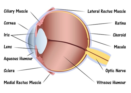
Anatomical structures of the human eye
External Structures
| Structure | Description | Function |
|---|---|---|
| Cornea | Transparent front surface | Primary refractive element (≈43D) |
| Sclera | White, fibrous outer layer | Structural support and protection |
| Conjunctiva | Thin, transparent membrane | Lubrication and protection |
Internal Structures
| Structure | Description | Function |
|---|---|---|
| Iris | Colored part with pupil | Controls light entry (aperture) |
| Lens | Transparent, biconvex structure | Fine focusing (accommodation) |
| Retina | Neural tissue with photoreceptors | Light detection and signal processing |
| Optic Nerve | Bundle of nerve fibers | Transmits visual information to brain |
Physiology of Vision
Optical System
The eye functions as a compound optical system with multiple refractive surfaces:
- Cornea-air interface (primary refractive power)
- Lens (variable focusing)
- Aqueous and vitreous humors (maintain shape and transmit light)
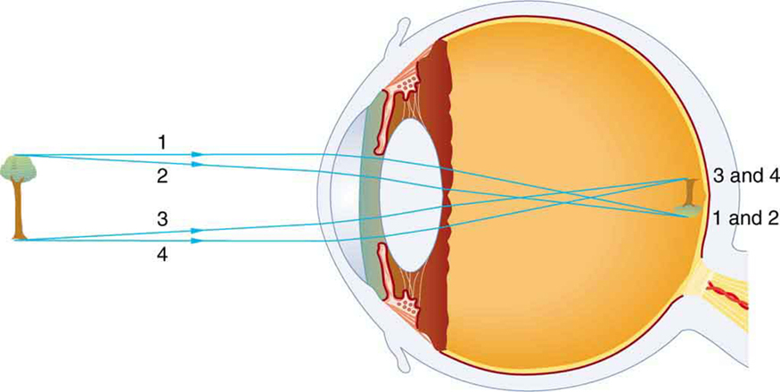
Figure: An image is formed on the retina with light rays converging most at the cornea and upon entering and exiting the lens. Rays from the top and bottom of the object are traced and produce an inverted real image on the retina.
Phototransduction
The process of converting light energy into neural signals:
- Light enters through cornea and pupil
- Lens focuses light onto retina
- Photoreceptors (rods and cones) capture photons
- Photopigment molecules undergo conformational change
- Neural signal generated and processed by retinal neurons
- Signal transmitted via optic nerve to visual cortex
Biomedical Engineering Perspective
Understanding the eye's optical properties is essential for designing intraocular lenses, contact lenses, laser vision correction systems, and retinal implants. The eye's approximate refractive power is 60D, with the cornea contributing about 43D and the lens about 17D.
Visual Pathway
The neural pathway from retina to visual cortex:
- Retina: Photoreceptors → bipolar cells → ganglion cells
- Optic Nerve: Axons of ganglion cells
- Optic Chiasm: Partial crossing of fibers
- Lateral Geniculate Nucleus (LGN): Thalamic relay station
- Optic Radiations: Pathway to visual cortex
- Primary Visual Cortex (V1): Initial cortical processing
Visual Processing Terms
- Visual Field: Total area visible without moving the eye
- Contralateral Processing: Left visual field processed by right hemisphere
- Receptive Field: Region of visual space that affects a neuron's activity